Embarking on an exploration of microscopic anatomy and organization of skeletal muscle exercise 12, this discourse delves into the intricate workings of skeletal muscle, providing a comprehensive understanding of its structure, adaptations, and functional implications.
Microscopic anatomy and organization play a pivotal role in skeletal muscle function during exercise. The hierarchical organization of skeletal muscle, from the whole muscle to the myofibril, dictates its contractile properties and response to exercise.
Introduction: Microscopic Anatomy And Organization Of Skeletal Muscle Exercise 12
The microscopic anatomy and organization of skeletal muscle play a crucial role in its function during exercise. Understanding these structures and their adaptations to exercise is essential for optimizing muscle performance and recovery.
Skeletal muscle is organized hierarchically, from the whole muscle to the myofibril. Each level of organization contributes to the overall contractile properties of the muscle.
Structure of Skeletal Muscle, Microscopic anatomy and organization of skeletal muscle exercise 12
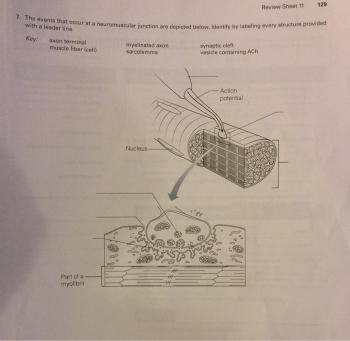
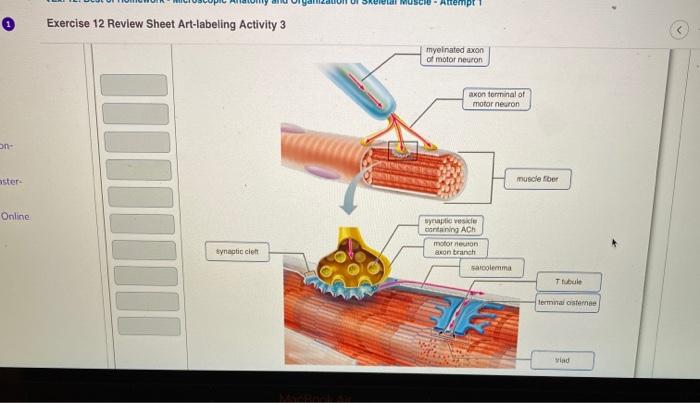
The sarcolemma is the outermost layer of the muscle fiber, which maintains the integrity of the cell and regulates the exchange of substances between the muscle fiber and the extracellular environment.
The sarcoplasm is the cytoplasm of the muscle fiber, which contains the myofibrils, mitochondria, and other organelles necessary for muscle function.
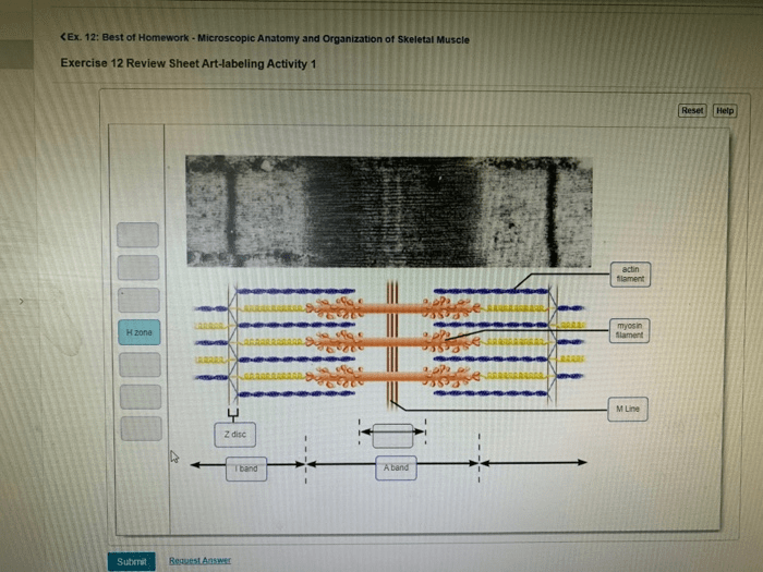
Myofibrils are the contractile units of the muscle fiber. They are composed of repeating units called sarcomeres, which contain the actin and myosin myofilaments that generate force.
The sarcoplasmic reticulum is a network of tubules that surrounds the myofibrils. It stores calcium ions, which are released during muscle contraction.
Transverse tubules are invaginations of the sarcolemma that penetrate the muscle fiber. They allow the rapid spread of electrical signals throughout the muscle fiber, triggering muscle contraction.
Microscopic Adaptations to Exercise
Exercise training can induce a variety of microscopic adaptations in skeletal muscle, including:
- Increased myofibril size and number
- Increased mitochondrial density
- Increased capillary density
- Changes in muscle fiber type composition
These adaptations enhance muscle function by increasing strength, power, and endurance.
Functional Implications
The microscopic anatomy and organization of skeletal muscle influence its contractile properties, including force, velocity, and power.
Muscle fiber type composition also affects muscle function. Type I fibers are slow-twitch and fatigue-resistant, while Type II fibers are fast-twitch and more powerful but fatigue more quickly.
Understanding microscopic anatomy can inform exercise prescription and recovery strategies. For example, endurance training favors adaptations that increase mitochondrial density and capillary density, while strength training favors adaptations that increase myofibril size and number.
FAQ Overview
What is the significance of microscopic anatomy in skeletal muscle function during exercise?
Microscopic anatomy provides insights into the structural organization of skeletal muscle, including the arrangement of myofilaments within the sarcomere and the role of the sarcoplasmic reticulum and transverse tubules in muscle contraction.
How does skeletal muscle adapt to different types of exercise training?
Skeletal muscle adapts to exercise training by altering its microscopic structure, such as increasing the number of mitochondria and capillaries, and modifying the composition of muscle fiber types.
What is the role of satellite cells in muscle growth and repair?
Satellite cells are stem cells that reside on the surface of muscle fibers and play a crucial role in muscle growth and repair by differentiating into new muscle fibers.